Concurrent malasseziosis and otitis externa in a senior pug

Downloads
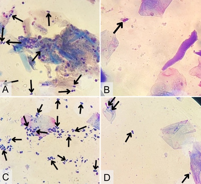
Dermatitis is a common skin disorder in dogs, caused by a variety of factors including fungi, ectoparasites, bacteria, and metabolic diseases. This case report describes a 7-year-old male pug with dermatitis associated with Malassezia infection and concurrent otitis externa caused by bacterial and Malassezia infections. The dog exhibited clinical signs, including pruritus, erythema, scaling, hyperpigmentation, lichenification, malodor, oily coat, and wet, malodorous ear discharge. Cytological analysis of skin and ear samples, performed using acetate tape preparation and otic swabs, revealed the presence of Malassezia yeast and cocci-shaped bacteria. Hematological evaluation revealed normocytic normochromic anemia. Based on these findings, the dog was diagnosed with Malasseziosis and bacterial-Malassezia otitis externa. A comprehensive therapeutic regimen was implemented, including oral antifungal ketoconazole, sebazole shampoo, oticon ear drops, anti-inflammatory methylprednisolone, antihistamine chlorpheniramine maleate, and supportive supplementation with multivitamins (Livron B-plex) and fish oil. After 21 days of treatment, significant clinical improvement was observed, demonstrating the efficacy of this multimodal therapeutic approach.
Cabañes FJ. 2021. Diagnosis of Malassezia Dermatitis and otitis in dogs and cats, is it just a matter of counting? Revista Iberoamericana de Micologia. 38(1):3-4. https://doi.org/10.1016/j.riam.2020.03.001 | PMid:32448732
Çınar M, Yağcı BB. 2021. Determination of Malassezia spp. infection and flea allergy incidences in pet dogs found in Kırıkkale and Ankara regions. Turkish Journal of Veterinary Research. 5(2):81-88. https://doi.org/10.47748/tjvr.953086
Ernawati, M, Soma, IG, Suarha, IN. 2023. Laporan kasus: demodekosis disertai dermatitis akibat infeksi jamur Malassezia Sp. pada anjing Shih Tzu. Jurnal Buletin Veteriner Udayana. 15 (3):410-422. https://doi.org/10.24843/bulvet.2023.v15.i03.p09
Guillot J, Bond R. 2020. Malassezia yeasts in veterinary dermatology: an updated overview. Frontiers in Cellular and Infection Microbiol-ogy.10(79):1-1. https://doi.org/10.3389/fcimb.2020.00079 | PMid:32181160 PMCid:PMC7059102
Purnama KA, Winaya IBO, Adi AAAM, Erawan IGMK, Kardena IM, Suartha IN. 2019. Gambaran histopatologi kulit anjing penderita dermatitis. Jurnal Veteriner. 20(4):486-496.
Seetha U, Kumar S, Pillai RM, Srinivas MV, Antony PX, Mukhopadhyay HK. 2018. Malassezia species associated with dermatitis in dogs and their antifungal susceptibility. International Journal of Current Microbiology and Applied Sciences. 7(6):1994-2007. https://doi.org/10.20546/ijcmas.2018.706.236
Sudipa PH, Gelgel KTP, Jayanti PD. 2021. Malassezia sp. infection prevalence in dermatitis dogs in Badung area. Advances in Tropical Biodiversity and Environmental Sciences. 5(2):45-49. https://doi.org/10.24843/ATBES.2021.v05.i02.p02
Ulfa Z, Elfidasari D, Sugoro I. 2016. Identifikasi khamir patogen pada kulit dan telinga anjing peliharaan. Jurnal Al-Azhar Indonesia Seri Sains dan Teknologi. 3(4):213-222. https://doi.org/10.36722/sst.v3i4.236
Sharma P, Rana T. 2023. Fungal diseases of goats. Principles of Goat Disease and Prevention. 111-126. https://doi.org/10.1002/9781119896142.ch9
Copyright (c) 2025 CC-BY-SA

This work is licensed under a Creative Commons Attribution-ShareAlike 4.0 International License.
All articles published in this journal are licensed under a Creative Commons Attribution-ShareAlike 4.0 International License (CC BY-SA 4.0).
This license permits use, distribution, reproduction, and adaptation in any medium, including for commercial purposes, provided the original work is properly cited, a link to the license is given, and any changes made are indicated. Any derivative work must be distributed under the same license or a compatible license.
How to Cite