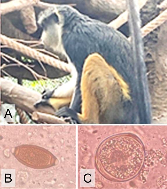
Helminthic and protozoan coinfections in mona monkeys (Cercopithecus mona) at Batu Secret Zoo, Batu, East Java, Indonesia
Parasitic coinfection in monkey

Downloads
This case report originated from fecal examination of six mona monkeys, which lived together in a cage. The helminth eggs from the feces were evaluated microscopically using the native method. Six eggs of Trichuris sp. were found, and the monkeys were subsequently treated with Curcuma Plus® orally for 3 days, combined by 3 days of fenbendazole orally. On the 6th day, no Trichuris sp. eggs were found; however, Entamoeba sp. cysts were discovered. No Entamoeba sp. cysts were identified on day 13. Helminthic infections will frequently dominate during protozoal and helminthic co-infection stages. A protozoal infection can be recognized after the helminth infection, which was treated with anthelmintics.
Boroumand N, Samarghandian S, Hashemy SI. 2018. Immunomodulatory, anti-inflammatory, and antioxidant effects of curcumin. Journal of Herbmed Pharmacology. 7(4):211-219. https://doi.org/10.15171/jhp.2018.33 DOI: https://doi.org/10.15171/jhp.2018.33
Dogra N, Kumar A, Mukhopadhyay T. 2018. Fenbendazole acts as a moderate microtubule destabilizing agent and causes cancer cell death by modulating multiple cellular pathways. Scientific reports. 8(1):11926. https://doi.org/10.1038/s41598-018-30158-6 | PMid:30093705 PMCid:PMC6085345 DOI: https://doi.org/10.1038/s41598-018-30158-6
Hailu GG, Ayele E T. 2021. Assessment of the prevalence of intestinal parasitic infections and associated habit and culture-related risk fac-tors among primary schoolchildren in Debre Berhan town, Northeast Ethiopia. BMC Public Health. 21(1): 1-12. https://doi.org/10.1186/s12889-020-10148-y | PMid:33422051 PMCid:PMC7797111 DOI: https://doi.org/10.1186/s12889-020-10148-y
Li M, Zhao B, Li B, Wang Q, Niu L, Deng J, Gu X, Peng X, Wang T, Yang G. 2015. Prevalence of gastrointestinal parasites in captive non‐human primates of twenty‐four zoological gardens in China. Journal of Medical Primatology. 44(3):168-173. https://doi.org/10.1111/jmp.12170 | PMid:25851745 PMCid:PMC6680269 DOI: https://doi.org/10.1111/jmp.12170
Mbuthia P, Murungi E, Owino V, Akinyi M, Eastwood G, Nyamota R, Lekolool I, Jeneby M. 2021. Potentially zoonotic gastrointestinal nematodes co‐infecting free ranging non‐human primates in Kenyan urban centres. Veterinary Medicine and Science. 7(3):1023-33. https://doi.org/10.1002/vms3.424 | PMid:33400394 PMCid:PMC8136933 DOI: https://doi.org/10.1002/vms3.424
Murphy H. 2015. Great Apes in Zoo and Wild Animal Medicine, Vol. 8, Miller RE, Fowler ME, Editors. Elsevier Saunders: St.Louis, Missouri. https://doi.org/10.1016/B978-1-4557-7397-8.00038-4 DOI: https://doi.org/10.1016/B978-1-4557-7397-8.00038-4
Riswanda Z, Kurniawan B. 2016. Infeksi soil-transmitted helminth: ascariasis, trichuriasis, dan cacing tambang. Majority. 5:61-68.
Sultana T, Jan U, Lee H, Lee H, Lee JI. 2022. Exceptional Repositioning of Dog Dewormer: Fenbendazole Fever. Current Issue in Molec-ular Biology. 44:4977-4986. https://doi.org/10.3390/cimb44100338 | PMid:36286053 PMCid:PMC9600184 DOI: https://doi.org/10.3390/cimb44100338
All articles published in this journal are licensed under a Creative Commons Attribution-ShareAlike 4.0 International License (CC BY-SA 4.0).
This license permits use, distribution, reproduction, and adaptation in any medium, including for commercial purposes, provided the original work is properly cited, a link to the license is given, and any changes made are indicated. Any derivative work must be distributed under the same license or a compatible license.
How to Cite