Cytological and complete blood count profile in a dog with suspected skin tumours

Downloads
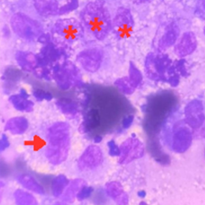
Skin tumours are among the most prevalent neoplasms in older dogs, and often display diverse clinical signs. This case report outlines the clinical presentation, diagnosis, and treatment of a suspected skin tumour in a 10-year-old obese female Golden Retriever weighing 46 kg. The owner brought the dog to West Java Provincial Animal Hospital with mandibular swelling, decreased appetite, and tachypnoea. Physical examination revealed a rectal temperature of 40.1°C, painful mandibular swelling, respiratory rate of 236 breaths per minute, and heart rate of 88 beats per minute. Cytology revealed pleomorphism, coarse chromatin, and nuclear moulding, suggesting increased cellular activity. Haematological results showed leukocytosis, granulocytosis, and hyperchromic normocytic anaemia, likely due to the release of proinflammatory cytokines from the tumour and immune cells. Differential diagnosis included sialadenitis, apocrine gland tumours, and salivary gland adenocarcinoma. The final diagnosis was skin tumour with poor prognosis. The treatment involved iron dextran, supplements, dexamethasone, sulfadiazine-trimethoprim, cyproheptadine HCl, and tolfenamic acid
Brazzell JL, Heinrich D, Walz JZ. 2020. Dermal and subcutaneous masses. Veterinary Cytology. 115-137. https://doi.org/10.1002/9781119380559.ch12 DOI: https://doi.org/10.1002/9781119380559.ch12
De Nardi AB, dos Santos Horta R, Fonseca-Alves CE, de Paiva FN, Linhares LC, Firmo BF, Ruiz Sueiro FA, de Oliveira KD, Lourenço SV, De Francisco Strefezzi R, Brunner CH. 2022. Diagnosis, prognosis and treatment of canine cutaneous and subcutaneous mast cell tumors. Cells. 11(4):618. https://doi.org/10.3390/cells11040618 | PMid:35203268 PMCid:PMC8870669 DOI: https://doi.org/10.3390/cells11040618
Filgueira KD, Chalita MC, Sellera FP, Reche Junior A. 2022. Cyto-pathology of cutaneous and subcutaneous neoplasms in feline species: a retrospective study. Acta Veterinaria Brasilica. 16(3):220-226. https://doi.org/10.21708/avb.2022.16.3.10717 DOI: https://doi.org/10.21708/avb.2022.16.3.10717
Hassan BB, Al-Mokaddem AK, Abdelrahman HA, Samir A, Mousa MR. 2022. Cutaneous tumors in dogs: a retrospective epidemiological and histological study of 112 cases. Advances in Animal and Veterinary Sciences. 10(1):170-182. https://doi.org/10.17582/journal.aavs/2022/10.1.170.182 DOI: https://doi.org/10.17582/journal.aavs/2022/10.1.170.182
Laskou S, Sapalidis K, Topalidis C, Koletsa T, Kesisoglou I. 2022. Be (a) ware of leukocytosis in papillary thyroid cancer. Case Reports in Endocrinology. 2022(1):5799432. https://doi.org/10.1155/2022/5799432 | PMid:35880158 PMCid:PMC9308557 DOI: https://doi.org/10.1155/2022/5799432
Madeddu C, Gramignano G, Astara G, Demontis R, Sanna E, Atzeni V, Macciò A. 2018. Pathogenesis and treatment options of cancer related anemia: perspective for a targeted mechanism-based approach. Frontiers in physiology. 9:1294. https://doi.org/10.3389/fphys.2018.01294 | PMid:30294279 PMCid:PMC6159745 DOI: https://doi.org/10.3389/fphys.2018.01294
Mango EE, Kardena IM, Supartika IK. 2016. Prevalensi dan gambaran histopatologi tumor kulit pada anjing di Kota Denpasar. Buletin Veteriner Udayana Volume. 8(1):65-70.
Mehrotra R, Singh M, Singh PA, Mannan R, Ojha VK, Singh P. 2007. Should fine needle aspiration biopsy be the first pathological investigation in the diagnosis of a bone lesion? An algorithmic approach with review of literature. CytoJournal. 4:9. https://doi.org/10.1186/1742-6413-4-9 | PMid:17439659 PMCid:PMC1872031 DOI: https://doi.org/10.1186/1742-6413-4-9
Millrud CR, Månsson Kvarnhammar A, Uddman R, Björnsson S, Riesbeck K, Cardell LO. 2012. The activation pattern of blood leukocytes in head and neck squamous cell carcinoma is correlated to survival. PloS one. 7(12):e51120. https://doi.org/10.1371/journal.pone.0051120 | PMid:23251433 PMCid:PMC3519486 DOI: https://doi.org/10.1371/journal.pone.0051120
Polymeris A, Kogia C, Ioannidis D, Lilis D, Drakou M, Maounis N, Kaklamanis L, Tseleni-Balafouta S. 2020. Excessive leukocytosis leading to a diagnosis of aggressive thyroid anaplastic carcinoma: a case report and relevant review. European Thyroid Journal. 9(3):162-168. https://doi.org/10.1159/000506767 | PMid:32523893 PMCid:PMC7265710 DOI: https://doi.org/10.1159/000506767
Putranto G, Bhaskara I, Batan I, Soma I. 2022. Laporan kasus: Metastasis ekstragenital tumor kelamin menular pada anjing peranakan pomeranian jantan. Indonesia Medicus Veterinus. [Online]. 11(3):371-385. https://doi.org/10.19087/imv.2022.11.3.371 DOI: https://doi.org/10.19087/imv.2022.11.3.371
Salvi M, Molinari F, Iussich S, Muscatello LV, Pazzini L, Benali S, Banco B, Abramo F, De Maria R, Aresu L. 2021. Histopathological classification of canine cutaneous round cell tumors using deep learning: a multi-center study. Frontiers in Veterinary Science. 8:640944. https://doi.org/10.3389/fvets.2021.640944 | PMid:33869320 PMCid:PMC8044886 DOI: https://doi.org/10.3389/fvets.2021.640944
All articles published in this journal are licensed under a Creative Commons Attribution-ShareAlike 4.0 International License (CC BY-SA 4.0).
This license permits use, distribution, reproduction, and adaptation in any medium, including for commercial purposes, provided the original work is properly cited, a link to the license is given, and any changes made are indicated. Any derivative work must be distributed under the same license or a compatible license.
How to Cite