Penanganan klinis dermatitis termal pada kucing domestik berbulu pendek.

Dermatitis termal adalah cedera kulit akibat luka bakar yang dapat menyebabkan kerusakan jaringan, peradangan, dan infeksi bakteri sekunder. Laporan kasus ini menggambarkan diagnosis, pengobatan, dan perkembangan penyembuhan dermatitis termal pada kucing domestik berbulu pendek. Pasien datang dengan dugaan luka bakar air panas di bagian tubuh kiri, dengan tanda-tanda klinis termasuk alopesia, eritema, eksudasi basah, dan keluaran nanah. Berdasarkan anamnesis dan pemeriksaan fisik, kasus ini didiagnosis sebagai dermatitis termal derajat dua yang diperumit oleh infeksi bakteri sekunder. Pengobatan meliputi antibiotik sistemik, terapi antiseptik dan antibiotik topikal, suplementasi vitamin, dan pembersihan luka rutin. Selama periode tindak lanjut delapan minggu, lesi menunjukkan perbaikan progresif, yang berpuncak pada penutupan luka yang lengkap, re-epitelisasi, dan pertumbuhan kembali rambut. Disimpulkan bahwa terapi multimodal yang konsisten secara efektif mencapai penyembuhan lengkap dalam kasus ini.
■ INTRODUCTION
Dermatitis is one of the most common dermatological disorders in veterinary practice, arising from parasitic, fungal, bacterial, and thermal causes. The skin serves as the body's protective barrier, maintaining homeostasis and preventing environmental injury. Direct exposure to external stressors makes it vulnerable to disruption. Thermal exposure is particularly significant, as it can cause tissue injury, inflammation, compromised barrier integrity, and an increased risk of secondary infection (Alautaish et al., 2024).
Thermal dermatitis occurs following contact with high temperature sources and manifests with varying severity, ranging from mild erythema to deep dermal destruction and full-thickness necrosis. Clinical outcomes depend on the injury depth, extent, and promptness of therapeutic intervention. If inadequately managed, thermal skin injuries may lead to complications, including bacterial contamination, delayed wound contraction, and compromised healing (Bond et al., 2020).
Given these complications, an early and accurate diagnosis is essential for appropriate case management and prognosis. Diagnostic evaluation requires a systematic approach, including anamnesis, physical examination, and assessment of lesion characteristics. Therapeutic management focuses on wound cleansing, infection control, inflammation reduction, and supportive measures for tissue regeneration (Wang et al., 2021). This case report describes the diagnostic workup and management of thermal dermatitis and documents the therapeutic response following veterinary intervention.
■ CASE
Signalment: A male domestic shorthair cat named Como with a black-and-white coat.
Anamnesis: The cat presented to Klinik Kumi Pet Store and Care with a history of suspected hot-water scald injury on the left side of the body and redness of the left eye.
Clinical Findings: Lesions on the left lateral body region included alopecia, erythema, moist exudative wounds, dull and broken hair, irregular wound margins, purulent discharge, and malodor. Conjunctival redness was also observed in the left eye.
Physical Examination: The findings were consistent with a partial-thickness thermal skin injury of moderate severity, without gross evidence of full thickness necrosis.
Laboratory Examination: No hematological or biochemical examinations were performed.
Differential Diagnosis: Thermal dermatitis, chemical dermatitis, traumatic dermatitis, and infectious dermatitis.
Diagnosis: Thermal dermatitis, consistent with a suspected second-degree burn, complicated by a secondary bacterial infection.
Prognosis: Fausta.
Treatment: Treatment included intramox LA 150 (15 mg/kg BW, IM, every 48 h), cefixime 100 mg (10 mg/kg BW, PO, BID), vitol 140 (0.5 mL/kg BW, PO, SID), aminoplex (0.5 mL/kg BW, PO, SID), and ADE fish oil (1 capsule, PO, SID). Topical therapy consisted of 0.9% NaCl irrigation, chlorhexidine cleansing, and Enbatic cream, with an e-collar application to prevent self-trauma.
■ RESULTS AND DISCUSSION
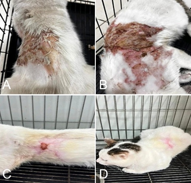
A male domestic cat presented with a thermal wound affecting the left dorsal body region, most likely associated with hot-water exposure, at the Kumi Pet Care Clinic based on anamnesis and physical examination. Clinically, the lesion was characterized by alopecia, erythema, moist exudation, and partial-thickness skin loss, findings compatible with second degree thermal dermatitis involving the epidermis and superficial dermis (Figure 1A and B).
Figure 1.Clinical presentation and healing of thermal dermatitis in a domestic short-haired cat. A-B. Initial presentation showing erythema, alopecia, and moist exudative skin injury. C-D. Wound healing from Week 1 to Week 8, showing reduced inflammation, wound contraction, and hair regrowth.
Thermal injury compromises the structural and protective functions of the epidermal barrier, predisposing the affected tissue to opportunistic bacterial contamination and secondary infection. In the present case, this was reflected by the appearance of purulent discharge, malodor, and crust formation, all of which are commonly associated with early wound contamination and active inflammation. These findings were considered indicative of the inflammatory phase of wound healing based on gross clinical assessment, although no hematological or biochemical examinations were performed. With continued treatment, the wound progressed into the proliferative phase, as demonstrated by the formation of healthy granulation tissue and early re-epithelialization, before ultimately entering the remodeling phase, which was characterized by complete wound closure and gradual hair regrowth.
The therapeutic regimen appeared to support each phase of wound repair. Systemic cefixime therapy likely contributed to bacterial control, thereby limiting inflammatory extension and reducing the risk of further tissue damage (Omrani et al., 2025). Meanwhile, topical wound care using chlorhexidine and enbatic cream helped reduce the superficial microbial burden and maintain a favorable local healing environment. Oral supplementation with Vitol 140 and Aminoplex may also have supported tissue recovery through improved nutritional status, immune support, and collagen synthesis. In addition, routine irrigation with 0.9% NaCl facilitated the removal of wound debris and exudate, while the application of an Elizabethan collar (e-collar) played an important role in preventing self-inflicted trauma and preserving wound stability throughout the healing process (Pournamdari et al., 2019).
Clinically, the wound exhibited a favorable healing trajectory. By the second week, there was a marked reduction in inflammation and exudation. By the fourth week, granulation tissue and re-epithelialization were clearly visible, indicating active tissue regeneration. By weeks 7-8, the wound had undergone complete closure, accompanied by early hair regrowth, consistent with advanced remodeling (Figure 1C and D).
■ CONCLUSION
This case demonstrates that early diagnosis, appropriate infection control, and consistent multimodal therapy are essential for the successful management of thermal dermatitis in cats. The complete recovery observed within eight weeks is in accordance with the expected healing course of partial thickness thermal injuries under appropriate veterinary care.
Referensi
- Alautaish H., Naji H.A., Saud Z.. Clinical study of common bacterial, fungal and parasitic skin diseases in cats. Advancements in Life Sciences. 2024; 11(3)DOI
- Bond R., Morris D.O., Guillot J., Bensignor E.J., Robson D., Mason K.V., Kano R., Hill P.B.. Biology, diagnosis and treatment of Malassezia dermatitis in dogs and cats: Clinical consensus guidelines of the world association for veterinary dermatology. Veterinary Dermatology. 2020; 31(1)DOI
- Omrani Z., Pourmadadi M., Rahdar A., Ghotekar S.. Recent advances in cefixime-loaded nanomaterials for treating bacterial infections. BioNanoScience. 2025; 15(3)DOI
- Pournamdari A.B., Tkachenko E., Barbieri J., Adamson A.S., Mostaghimi A.. A state-of-the-art review highlighting medical overuse in dermatology, 2017-2018: A systematic review. JAMA Dermatology. 2019; 155(12)DOI
- Wang V., Boguniewicz J., Boguniewicz M., Ong P.Y.. The infectious complications of atopic dermatitis. Asthma & Immunology. 2021; 126(1)DOI
Alautaish HHN, Naji HA, Saud ZAH. 2024. Clinical study of common bacterial, fungal and parasitic skin diseases in cats. Advancements in Life Sciences. 11(3):580-584. https://doi.org/10.62940/als.v11i3.1873
Bond R, Morris DO, Guillot J, Bensignor EJ, Robson D, Mason KV, Kano R, Hill PB. 2020. Biology, diagnosis and treatment of Malassezia dermatitis in dogs and cats: Clinical consensus guidelines of the world association for veterinary dermatology. Veterinary Dermatology. 31(1):75. https://doi.org/10.1111/vde.12834 | PMid:31957203
Omrani Z, Pourmadadi M, Rahdar A, Ghotekar S. 2025. Recent advances in cefixime-loaded nanomaterials for treating bacterial infections. BioNanoScience. 15(3):467. https://doi.org/10.1007/s12668-025-02087-y
Pournamdari AB, Tkachenko E, Barbieri J, Adamson AS, Mostaghimi A. 2019. A state-of-the-art review highlighting medical overuse in dermatology, 2017-2018: A systematic review. JAMA Dermatology. 155(12):1410-1415. https://doi.org/10.1001/jamadermatol.2019.3064 | PMid:31642872
Wang V, Boguniewicz J, Boguniewicz M, Ong PY. 2021. The infectious complications of atopic dermatitis. Annals of Allergy, Asthma & Immunology. 126(1):3-12. https://doi.org/10.1016/j.anai.2020.08.002 | PMid:32771354 PMCid:PMC7411503
Hak Cipta (c) 2026 CC-BY-SA

Artikel ini berlisensiCreative Commons Attribution-ShareAlike 4.0 International License.
Karya ini dilisensikan di bawah Creative Commons Attribution-ShareAlike 4.0 International License (CC BY-SA 4.0).
Lisensi ini mengizinkan siapa pun untuk menyalin, membagikan, menggunakan kembali, mengadaptasi, mengubah, dan mengembangkan karya ini dalam media atau format apa pun, termasuk untuk tujuan komersial, dengan syarat mencantumkan atribusi yang sesuai kepada penulis/pencipta asli, menyertakan tautan ke lisensi, dan menjelaskan jika ada perubahan yang dilakukan.
Apabila karya ini diadaptasi atau dimodifikasi, hasil adaptasinya harus didistribusikan dengan lisensi yang sama atau lisensi yang kompatibel.
Cara Mengutip