Renal lipid peroxidation and tubular lesions following ethylene glycol and ammonium chloride exposure in rats

Downloads
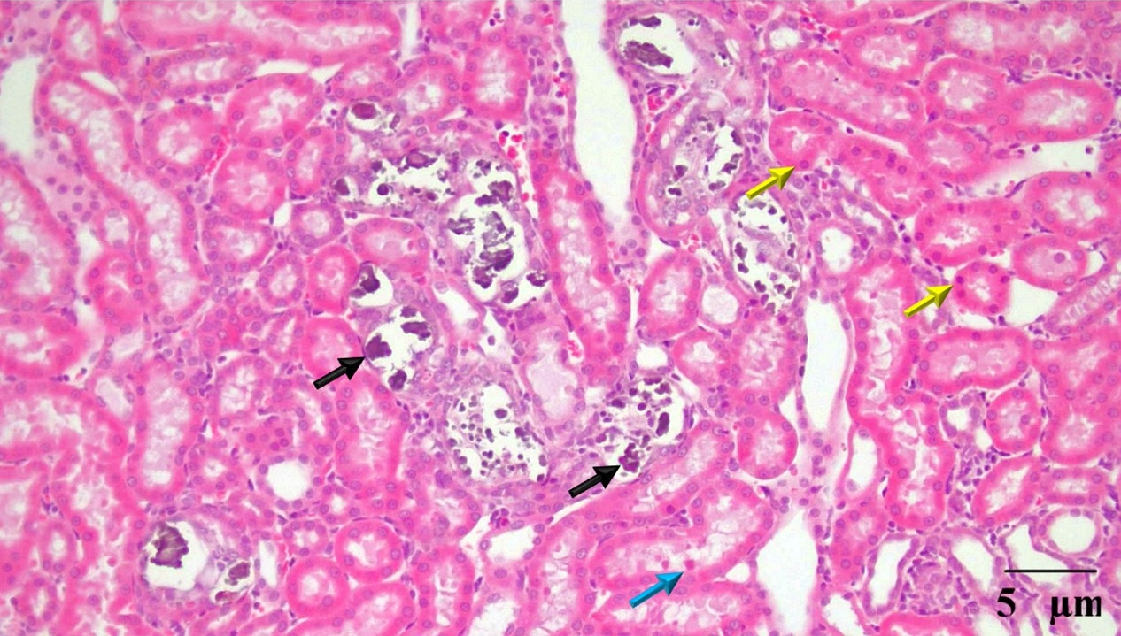
Nephrolithiasis-induced renal injury involves crystal deposition and oxidative stress. This study evaluated renal malondialdehyde (MDA) levels and histopathological alterations in rats induced with ethylene glycol (EG) and ammonium chloride (NH₄Cl). Ten male Sprague–Dawley rats were assigned to a normal control group and an induction group receiving 0.75% EG and 2% NH₄Cl in drinking water ad libitum for 28 days (n = 5 per group). Renal MDA concentrations were quantified via thiobarbituric acid reactive substances (TBARS) assay, and tissue structural changes were evaluated using hematoxylin and eosin staining. The induction group exhibited a statistically significant elevation in renal MDA levels (11.12 ± 4.45 nmol/mg wet tissue) compared to controls (5.93 ± 0.61 nmol/mg wet tissue). Histopathological examination revealed intratubular crystal deposition, hyaline cast formation, and tubular epithelial cell necrosis. Co-administration of EG and NH₄Cl effectively establishes a nephrolithiasis model characterized by heightened lipid peroxidation and severe nephron structural damage.
Alford A, Furrow E, Borofsky M, Lulich J. 2020. Animal models of naturally occurring stone disease. Nature Reviews Urology. 17(12):691-705. https://doi.org/10.1038/s41585-020-00387-4 | PMid:33159170 PMCid:PMC9254658
Green ML, Hatch M, Freel RW. 2005. Ethylene glycol induces hyperoxaluria without metabolic acidosis in rats. American Journal of Physiology-Renal Physiology. 289(3):F536-F543. https://doi.org/10.1152/ajprenal.00025.2005 | PMid:15855660
Ito F, Sono Y, Ito T. 2019. Measurement and clinical significance of lipid peroxidation as a biomarker of oxidative stress: oxidative stress in diabetes, atherosclerosis, and chronic inflammation. Antioxidants. 8(3):72. https://doi.org/10.3390/antiox8030072 | PMid:30934586 PMCid:PMC6466575
Khan SR. 2013. Reactive oxygen species as the molecular modulators of calcium oxalate kidney stone formation: evidence from clinical and experimental investigations. The Journal of Urology. 189(3):803-811. https://doi.org/10.1016/j.juro.2012.05.078 | PMid:23022011
Li X, Chen S, Feng D, Fu Y, Wu H, Lu J, Bao J. 2021. Calcium-sensing receptor promotes calcium oxalate crystal adhesion and renal injury in Wistar rats by promoting ROS production and subsequent regulation of PS ectropion, OPN, KIM-1, and ERK expression. Renal Failure. 43(1):465-476. https://doi.org/10.1080/0886022X.2021.1881554 | PMid:33678127 PMCid:PMC7946014
Mulay SR, Kulkarni OP, Rupanagudi KV, Migliorini A, Darisipudi MN, Vilaysane A, Muruve D, Shi Y, Munro F, Liapis H, Anders HJ. 2012. Calcium oxalate crystals induce renal inflammation by NLRP3-mediated IL-1β secretion. The Journal of Clinical Investigation. 123(1):236-246. https://doi.org/10.1172/JCI63679 | PMid:23221343
Prabhu VV, Sathyamurthy D, Ramasamy A, Das S, Anuradha M, Pachiappan S. 2016. Evaluation of protective effects of diosmin (a citrus flavonoid) in chemical-induced urolithiasis in experimental rats. Pharmaceutical Biology. 54(9):1513-1521. https://doi.org/10.3109/13880209.2015.1107105 | PMid:26799954
Purwono RM, Wientarsih I, Widodo S, Purwaningsih EH, Harlina E. 2019. Gambaran kristal urin pada hewan model nefrolitiasis hasil induksi etilen glikol. ARSHI Veterinary Letters. 3(1):19-20. https://doi.org/10.29244/avl.3.1.19-20
Tsikas D. 2017. Assessment of lipid peroxidation by measuring malondialdehyde (MDA) and relatives in biological samples: Analytical and biological challenges. Analytical Biochemistry. 524:13-30. https://doi.org/10.1016/j.ab.2016.10.021 | PMid:27789233 PMCid:PMC9536629
Zhu S, Wang Q, Sun F, Jiang K. 2024. Baicalin attenuated oxidative stress and inflammation in ethylene glycol-induced urolithiasis in adult male SD rats. Tissue and Cell. 89:102453. https://doi.org/10.1016/j.tice.2024.102453 | PMid:38964085
Copyright (c) 2026 Retno Wilujeng, Eva Harlina, Rini Madyastuti Purwono, Dimas Andrianto

This work is licensed under a Creative Commons Attribution-ShareAlike 4.0 International License.
All articles published in this journal are licensed under a Creative Commons Attribution-ShareAlike 4.0 International License (CC BY-SA 4.0).
This license permits use, distribution, reproduction, and adaptation in any medium, including for commercial purposes, provided the original work is properly cited, a link to the license is given, and any changes made are indicated. Any derivative work must be distributed under the same license or a compatible license.