Hematobiochemical comparison of fat-tailed sheep naturally infected with nematodes in the highlands
DOI:
https://doi.org/10.29244/avl.10.1.27-28Keywords:
Trichostrongylus sp, Strongyloides sp, highland, fat-tailed sheep, blood profileAbstract
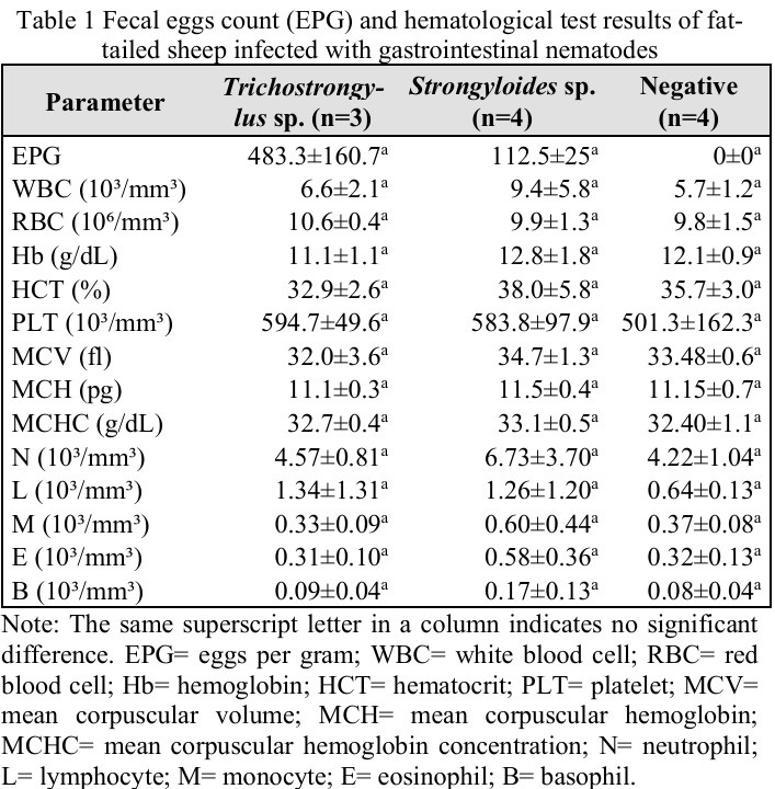
Gastrointestinal (GI) parasites are a major factor in reducing the productivity of small ruminants, particularly fat-tailed sheep, in tropical regions. This study aimed to evaluate the hematological and biochemical profiles of blood as indicators of GI nematode infection in sheep raised in the highlands. A total of 11 two-year-old ewes were used, consisting of three Trichostrongylus sp.-positive, four Strongyloides sp.-positive, and four negative ewes based on fecal examination. Hematological parameters were analyzed using ABX Micros 60, and biochemical parameters were analyzed using ABX Pentra C200. Data were analyzed using the Kruskal-Wallis test with Bonferroni's post hoc test. The results showed no significant differences (P>0.05) in any of the hematological and biochemical parameters between the infected and uninfected groups. This variation in hematopoietic responses is thought to be influenced by infection intensity, nutritional status, individual variation, sample size limitations, and environmental conditions
References
Alkateb YNM, Abdullah DA, Alobaidy AAA. 2023. Prevalence and haematobiochemical alterations associated with Strongyloides papillosus infection among Awassi breed of sheep in Mosul, Iraq. Comparative Clinical Pathology. 32: 225-230. https://doi.org/10.1007/s00580-022-03430-5
Charlier J, Rinaldi L, Musella V, Ploeger, HW, Chartier C, Vineer, HR. 2020. Initial assessment of the economic burden of major parasitic helminth infections to the ruminant livestock industry in Europe. Preventive Veterinary Medicine. 182:105103. https://doi.org/10.1016/j.prevetmed.2020.105103 | PMid:32750638
Dias-Silva T, Filho A, Katiki L, Amarante A, Abdalla A, Louvandini H. 2020. Trichostrongylus colubriformis infection in Santa Inês lambs: impact on feed digestibility, blood markers, and nitrogen balance. Revista Brasileira de Parasitologia Veterinaria. 29(2):1-7. https://doi.org/10.1590/s1984-29612020026 | PMid:32428186
Fernandes MA, Lima P, Amarante A, Abdalla A, Louvandini H. 2022. Hematological, biochemical alterations and methane production in sheep submitted to mixed infection of Haemonchus contortus and Trichostrongylus colubriformis. Small Ruminant Research. 216: 106798. https://doi.org/10.1016/j.smallrumres.2022.106798
Islam A, Islam S, Ferdous J, Rahman M, Uddin M, Akter S, Rahman M, Hassan M. 2019. Diversity and prevalence of parasitic infestation with zoonotic potential in dromedary camel (Camelus dromedarius) and fat-tailed sheep (dhumba) in Bangladesh. Journal of Advanced Veterinary and Animal Research. 6(1):142-147. https://doi.org/10.5455/javar.2019.f324 | PMid:31453183 PMCid:PMC6702934
Mavrot F, Hertzberg H, Torgerson P. 2015. Effect of gastrointestinal nematode infection on sheep performance: a systematic review and metaanalysis. Parasites & Vectors. 2(8):557. https://doi.org/10.1186/s13071-015-1164-z | PMid:26496893 PMCid:PMC4619485
Moosa D, Hussien A, Hameed H, Hasan S. 2022. Diagnostic and hematological study in sheep infected with gastrointestinal nematode in Mosul city. Al-Anbar Journal of Veterinary Sciences. 15(1):29-33. https://doi.org/10.37940/AJVS.2022.15.1.4
Rophi AH. 2015. Identifikasi cacing parasit dan prevalensinya pada ternak kambing di Kelurahan Koya Barat, Distrik Muara Tami, Kota Jayapura, Provinsi Papua. Novae Guinea Jurnal Biologi. 6(2).
Zeryehun T. 2012. Helminthosis of sheep and goats in and around Haramaya, Southeastern Ethiopia. Journal of Veterinary Medicine and Animal Health. 4(3):48-55.
Downloads
Published
Issue
Section
License
Copyright (c) 2026 CC-BY-SA

This work is licensed under a Creative Commons Attribution-ShareAlike 4.0 International License.
All articles published in this journal are licensed under a Creative Commons Attribution-ShareAlike 4.0 International License (CC BY-SA 4.0).
This license permits use, distribution, reproduction, and adaptation in any medium, including for commercial purposes, provided the original work is properly cited, a link to the license is given, and any changes made are indicated. Any derivative work must be distributed under the same license or a compatible license.