Anoplasty for type III atresia ani with fistula complicated by megacolon in a puppy

Downloads
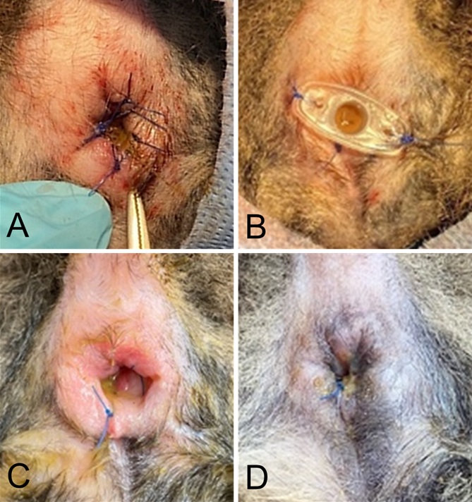
Congenital anorectal malformations, including atresia ani, are uncommon conditions in small animals that cause gastrointestinal dysfunction. This case report describes the surgical management and outcomes of type III atresia ani in a juvenile dog. A one-month-old female mixed-breed puppy presented with severe tenesmus, progressive abdominal distension, and complete absence of normal faecal passage. Clinical examination identified an anal dimple lacking a functional opening, accompanied by a small ventral perineal fistula. Radiographic assessment confirmed type III atresia ani, characterised by a blind-ending rectal pouch located more than 1 cm cranial to the anal dimple and associated with marked megacolon. Surgical intervention was performed via anoplasty, combined with manual evacuation of the impacted faeces and temporary anal stenting using a 1-cc syringe port to maintain luminal patency. Postoperative management included broad-spectrum antibiotics, lactulose, analgesic therapy, and meticulous wound care. Although normal defaecation was restored, the patient developed tenesmus, incomplete evacuation, and faecal incontinence. These complications are associated with irreversible megacolon and anal sphincter dysfunction. Conservative therapy failed to achieve improvement, indicating the need for surgical intervention.
Ettinger SJ, Feldman EC. 2010. Textbook of veterinary internal medicine (7th ed.). Philadelphia, PA: Saunders / Elsevier Health Sciences.
Ellison GW, Papazoglou LG. 2012. Long-term results of surgery for atresia ani with or without anogenital malformations in puppies and a kitten: 12 cases (1983-2010). Journal of the American Veterinary Medical Association. 240(2):186-192. https://doi.org/10.2460/javma.240.2.186 | PMid:22217027
Kim M, Hwang YH, Choi W, Lee JH. 2013. Surgical correction of congenital type III atresia ani with rectovaginal fistula in a cat. Journal of Veterinary Clinics. 30(5):376-379.
Prassinos NN, Papazoglou LG, Adamama‐Moraitou KK, Galatos AD, Gouletsou P, Rallis TS. 2003. Congenital anorectal abnormalities in six dogs. Veterinary Record. 153(3):81-85. https://doi.org/10.1136/vr.153.3.81 | PMid:12892267
Sharif S, Shaikh MY, Ali SM. 2019. Novel tool for minimally invasive brain surgery-Syringe port system. World Neurosurgery. 131:339-345. https://doi.org/10.1016/j.wneu.2019.06.202 | PMid:31284061
Copyright (c) 2025 CC-BY-SA

This work is licensed under a Creative Commons Attribution-ShareAlike 4.0 International License.
All articles published in this journal are licensed under a Creative Commons Attribution-ShareAlike 4.0 International License (CC BY-SA 4.0).
This license permits use, distribution, reproduction, and adaptation in any medium, including for commercial purposes, provided the original work is properly cited, a link to the license is given, and any changes made are indicated. Any derivative work must be distributed under the same license or a compatible license.
How to Cite