Luksasio patela pada kambing
Abstract
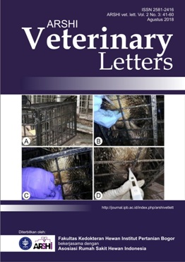
Luksasio persendian patela merupakan kondisi bawaan di mana tulang tempurung lutut (patela) mengalami dislokasi dari alur troklearis normal. Luksasio ini secara klinis dapat terjadi pada bagian medial, di dalam permukaan, lateral, maupun di luar permukaan lutut. Tulisan ini melaporkan kasus patela berpindah lokasi pada kambing. Tulang patela berpindah ke medial persendian lutut dan terlihat berada di belakang tulang femur, sehingga persendian lutut tidak dapat bergerak. Tindakan pembedahan dilakukan untuk melakukan reposisi. Insisi dimulai dari daerah parapatelar sekitar 1 cm dibagian lateral patela dan di atas sendi lutut. Jaringan sendi dipreparir dan dieksplorasi sehingga daerah femoro-tibia dapat diakses dengan baik. Inspeksi dan konfirmasi keadaan ligamen krusiate dari lutut dilakukan untuk melihat keparahan luksasio. Terdapat dua kelainan yang terjadi, yaitu troklea ossis femoris dangkal dan beberapa ligamen patela yang memendek karena kejadian dislokasi telah berlangsung kronis. Tindakan yang dilakukan adalah memperdalam cekungan pada troklea ossis femoris menggunakan kikir sebagai tempat pertautan patela. Selanjutnya melakukan pemotongan tendon (ligamen patela lateral) dan mereposisi patela. Berdasarkan citra radiografi menampakkan tulang patela berada di kranial tulang femur sehingga persendian lutut dapat bergerak dengan baik.
Downloads
References
Dyce KM, Sack WO, Wensing CJG. 2010. Textbook of Veterinary Anatomy. 4th Ed. Philadelpia (US): WB Saunders.
Harasen G. 2006a. Patellar luxation. CVJ. 47: 817-818.
Harasen G. 2006b. Patellar luxation: Pathogenesis and surgical correction. CVJ. 47: 1037-1039.
Isaka M, Befu M, Mastubara N, Ishikawa M, Arae Y, Tsuyama T, Doi A, Namba S. 2014. Corrective surgery for canine patellar luxation in 75 cases (107 limbs): Landmark for block reces-sion. Vet Sci Dev. 4(5251):30-32.
Johnson AL, Probst CW, Decamp CE, Rosenstein DS, Hauptman JG, Weaver BT, Kern TL. 2001. Comparison of trochlear block recession and trochlear wedge recession for canine patel-lar luxation using a cadaver model. Vet Surg. 30:140-150.
Copyright (c) 2018 CC-BY-SA

This work is licensed under a Creative Commons Attribution-ShareAlike 4.0 International License.
Authors who publish with this journal agree to the following terms:
1. Authors retain copyright and grant the journal right of first publication with the work simultaneously licensed under a Creative Commons Attribution License that allows others to share the work with an acknowledgement of the work's authorship and initial publication in this journal.
2. Authors are able to enter into separate, additional contractual arrangements for the non-exclusive distribution of the journal's published version of the work (e.g., post it to an institutional repository or publish it in a book), with an acknowledgement of its initial publication in this journal.
3. Authors are permitted and encouraged to post their work online (e.g., in institutional repositories or on their website) prior to and during the submission process, as it can lead to productive exchanges, as well as earlier and greater citation of published work (See The Effect of Open Access).


.jpg)


