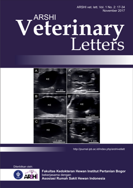
Pencitraan Ultrasonografi Organ Hepatobiliari pada Ular Sanca
Abstract
Teknologi pencitraan ultrasonografi memiliki peranan penting dalam manajemen kesehatan pada ular sanca. Struktur sistem organ di dalam tubuh seperti hepatobiliari dapat dipantau secara non-invasif menggunakan teknologi ultrasonografi. Citra ultrasonografi organ hepatobiliari pada tiga spesies ular sanca, yang terdiri atas sanca batik, sanca bodo, dan sanca bola dilaporkan dalam tulisan ini. Pencitraan organ hepatobiliari dilakukan menggunakan trasnduser linear dengan frekuensi 10 MHz. Ukuran organ hepatobiliari diukur berdasarkan nomor dan jumlah sisik ventral tubuh. Hasil pengukuran menunjukkan bahwa organ hati pada ketiga sanca memiliki ukuran yang bervariasi dan berada di antara persinggungan nomor sisik ventral 72-102. Organ kelenjar empedu berukuran 7-9 sisik dan berada di antara nomor sisik ventral yang bervariasi. Derajat ekogenitas organ hepatobiliari di antara ketiga sanca tidak menunjukkan adanya perbedaan. Ekogenisitas organ hati pada ketiga sanca tampak hipoekoik dengan dinding hiperekoik di antara bayangan organ paru-paru yang tampak hiperekoik. Kelenjar empedu tampak anekoik dengan dinding hiperekoik berada diantara organ limpa yang tampak hiperekoik dan organ pankreas yang tampak hipoekoik.Downloads
References
Banzato T, Russo E, Finotti L, Milan M, Gianesella M, Zotti A. 2012. Ultrasonographic anatomy of the coelomic organs of boid snakes. Am. J. Vet. Res. 73(5) 634-645.
Boyer JL. 2013. Bile formation and secretion. Compr. Physiol. 3(3): 1035-1078.
Gnudi G, Volta A, Ianni F, Bonazzi M, Manfredi S, Bertoni G. 2009. Use of ultrasonography and contrast radiography for snake gender determination. Vet. Radiol. Ultrasound. 50(3): 309-311.
Isaza R, Ackerman N, Jacobson ER. 1993. Ultrasound imaging of the coelomic structures in the Boa constrictor (Boa constrictor). Vet. Radiol. 34(6): 445-450.
Stahlschmidt Z, Brashears J, DeNardo D. 2011. The use of ultrasonography to assess reproductive investment and output in pythons. Biol. J. Linn. Soc. 103: 772-778.
Konde LJ, Pugh CR. 1996. Radiology and sonography of the digestive system. Dalam: Handbook of Small animal Gastroenterology. TR Tams, Ed. Philadelphia (US): Saunders.
Copyright (c) 2017 ARSHI Veterinary Letters

This work is licensed under a Creative Commons Attribution-ShareAlike 4.0 International License.
Authors who publish with this journal agree to the following terms:
1. Authors retain copyright and grant the journal right of first publication with the work simultaneously licensed under a Creative Commons Attribution License that allows others to share the work with an acknowledgement of the work's authorship and initial publication in this journal.
2. Authors are able to enter into separate, additional contractual arrangements for the non-exclusive distribution of the journal's published version of the work (e.g., post it to an institutional repository or publish it in a book), with an acknowledgement of its initial publication in this journal.
3. Authors are permitted and encouraged to post their work online (e.g., in institutional repositories or on their website) prior to and during the submission process, as it can lead to productive exchanges, as well as earlier and greater citation of published work (See The Effect of Open Access).


.jpg)


