Presumptive feline hemotropic mycoplasmosis in a domestic cat with anemia, thrombocytopenia, and epistaxis

Downloads
Feline hemotropic mycoplasmosis is a significant vector-borne disease in cats, causing anaemia and systemic illness. A 3-year-old male domestic cat (3.9 kg) showed decreased appetite, respiratory signs, and epistaxis. Despite complete vaccination, ectoparasite and anthelmintic control were inconsistent. Examination revealed a body condition score of 3/5, 7% dehydration, pale mucous membranes, prolonged capillary refill, hyperthermia (40 °C), and flea infestation. Haematology revealed leukopenia, lymphopenia, granulocytopenia, thrombocytopenia, and normocytic normochromic anaemia. The FPV test was negative. Blood smears showed organisms consistent with hemotropic Mycoplasma spp. The cat was diagnosed with presumptive feline hemotropic mycoplasmosis. Treatment included doxycycline (10 mg/kg PO q24h), tranexamic acid (0.1 mL/kg IM), aminophylline (20 mg/kg PO), Ornipural (5 mL for 5 days), and supportive therapy with vitamin B complex and iron. After 17 days of hospitalisation, the cat's appetite, activity, and haematological parameters improved significantly.
■ INTRODUCTION
Vector-borne diseases (VBDs) are arthropod-transmitted infections that remain a major cause of global morbidity and mortality, particularly in tropical and subtropical regions, where vector exposure is highly prevalent (Díaz-Regañón et al., 2018). Among feline VBDs, feline hemotropic mycoplasmosis is of particular clinical importance. This disease is caused by hemotropic Mycoplasma spp., which parasitise the surface of erythrocytes and are recognised as among the most pathogenic hemotropic bacterial infections in cats. In affected animals, untreated infection may progress to severe haemolytic anaemia and can ultimately be fatal (Tasker, 2018). The clinical presentation is often variable and is largely influenced by the host immune response, with commonly reported signs including pale mucous membranes, pyrexia, lethargy, anorexia, tachycardia, cardiac murmurs, weight loss, hepatomegaly, splenomegaly, lymphadenomegaly, and different degrees of haemolytic anaemia (Strandberg et al., 2023). Despite the recognised susceptibility of cats to this infection, clinically documented cases of feline hemotropic mycoplasmosis-particularly those accompanied by haemolytic anaemia -remain relatively limited in the literature. Consequently, additional well-documented clinical reports are needed to broaden the current understanding of disease presentation and to support improvements in diagnostic and therapeutic approaches.
■ CASE
Signalment and anamnesis: A 3-year-old male cat (3.9 kg) was presented with decreased appetite, flu-like signs, and epistaxis. Previous tests revealed thrombocytopenia. Vaccination was complete; however, ectoparasite control and deworming were inconsistent.
Physical examination: Pyrexia (40°C), respiratory rate of 32 breaths/min, heart rate of 120 beats/min, prolonged capillary refill time (>2 s), decreased skin turgor (>2 s), body condition score of 3/5, and pale mucous membranes.
Laboratory examinations: Haematological evaluation revealed leukopenia, lymphopenia, granulocytopenia, thrombocytopenia, and normocytic normochromic anaemia. FPV test was negative. Diff-Quik staining revealed hemotropic mycoplasma-like organisms on erythrocytes.
Differential diagnoses: feline panleukopenia, ehrlichiosis/anaplasmosis, FeLV-or FIV-associated haematologic disorders, immune-mediated haemolytic anaemia, coagulopathy, and other infectious or inflammatory anaemia causes.
Diagnosis: Presumptive feline haemotropic mycoplasmosis.
Prognosis: Favorable (fausta).
Treatment: Doxycycline (10 mg/kg PO q24 h), tranexamic acid (0.1 mL/kg IM), aminophylline (20 mg/kg PO), ornipural (5 mL once daily for 5 days), and supportive care.
| Parameters | Results | Normal range (Schalm et al., 2010) |
|---|---|---|
| White Blood Cells (×10³/µL) | 2.5 | 5.5–19.5 |
| Lymphocyte (%) | 17.0 | 12–45 |
| Granulocyte (%) | 78.5 | 35–85 |
| Monocyte (%) | 4.5 | 2–9 |
| Lymphocyte (×10³/µL) | 0.4 | 0.8–7.0 |
| Granulocyte (×10³/µL) | 2.0 | 2.1–15.0 |
| Monocyte (×10³/µL) | 0.1 | 0.0–1.9 |
| Red Blood Cell (×10⁶/µL) | 4.9 | 4.6–10.0 |
| Hemoglobin (g/dL) | 7.1 | 9.3–15.3 |
| Hematocrit (%) | 23.4 | 28–49 |
| MCV (fL) | 48.0 | 39–52 |
| MCH (pg) | 14.5 | 13–21 |
| MCHC (g/dL) | 30.3 | 30–38 |
| RDW-CV (%) | 26.1 | 14–18 |
| RDW-SD (fL) | 41.1 | 20–80 |
| Platelet (×10³/µL) | 49.0 | 100–514 |
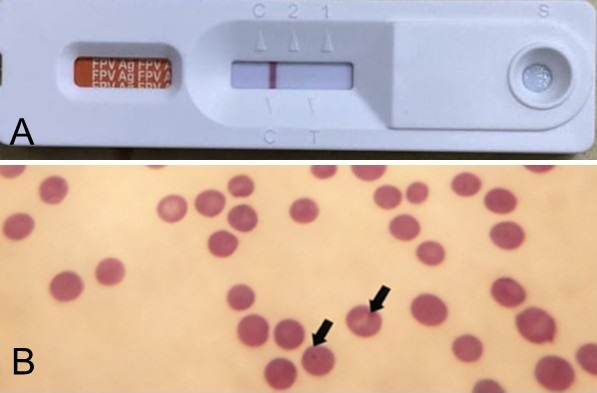
Figure 1.Laboratory findings in a 3-year-old male cat with presumptive feline hemotropic mycoplasmosis. (A) FPV rapid test result. (B) Diff-Quik-stained blood smear showing Mycoplasma-like organisms on erythrocytes (1000×).
■ RESULTS AND DISCUSSION
This report describes a 3-year-old cat with presumptive feline hemotropic mycoplasmosis, likely due to Mycoplasma haemofelis, associated with severe anaemia. Haematological abnormalities included leukopenia, lymphopenia, and granulocytopenia, suggesting a parasitic or bacterial infection ( Table 1). Decreased erythrocyte count, haemoglobin, and haematocrit levels indicated anaemia (Amelia et al., 2019). Although microcytic hypochromic anaemia is often linked to iron and copper deficiencies (Weiss & Wardrop, 2011), cytological and clinical findings supported this profile. The feline panleukopenia virus (FPV) rapid test was negative (Figure 1), making FPV-associated cytopenia unlikely. Blood smear evaluation showed bacterial organisms resembling Mycoplasma haemofelis attached to erythrocytes.
Definitive confirmation was not achieved as the diagnosis relied on blood smears and rapid testing, both with limited sensitivity. Thus, polymerase chain reaction (PCR), especially quantitative PCR (qPCR), is the gold standard for confirming feline hemotropic mycoplasma infection (Martínez-Díaz et al., 2013). Treatment focused on doxycycline as antimicrobial therapy, effective at oral doses of 5-10 mg/kg once daily (Purba et al., 2020)(Satriawan & Octaviani, 2021). Symptomatic therapy included tranexamic acid to minimise haemorrhage and aminophylline for bronchoconstriction. Ornipural was used as a hepatoprotective agent. Additional care included vitamin B complex, vitamin C, Sangobion, Neurobion, Hepaq, and Fu Fang (1 mL/day). Preventive management addressed vector-related infection through sanitation and ectoparasite control using Advocate with imidacloprid and moxidectin (Maslakah & Kusumarini, 2023). Clinical and haematological improvements were noted, with values normalising by day 15, and the cat was discharged healthy on day 17.
■ CONCLUSION
Early recognition and appropriate management of presumptive feline hemotropic mycoplasmosis may result in favourable clinical and haematological outcomes.
References
- Amelia F., Umbu D., Adistya E.R., Soesatyoratih R.R., Maylina L., Utami N.D., Cahyono T., Noviana D.. An integrative therapy for paraplegia dog with ehrlichiosis. ARSHI Veterinary Letters. 2019; 3(4)DOI
- Díaz-Regañón D., Villaescusa A., Ayllón T., Rodríguez-Franco F., García-Sancho M., Agulla B., Sainz Á.. Epidemiological study of hemotropic mycoplasmas (hemoplasmas) in cats from central Spain. Parasites & Vectors. 2018; 11(140)DOI
- Martínez-Díaz V.L., Silvestre-Ferreira A.C., Vilhena H., Pastor J., Francino O., Altet L.. Prevalence and co-infection of haemotropic mycoplasmas in Portuguese cats by real-time polymerase chain reaction. Journal of Feline Medicine and Surgery. 2013; 15(10)DOI
- Maslakah A.U., Kusumarini R.. Case study: Generalized demodicosis and malasseziosis in a pug mix dog. Journal of Parasite Science. 2023; 7(2)DOI
- Purba D.J., Widyastuti S.K., Anthara M.S.. Case report: Feline hemotropic mycoplasma in a local cat. Indonesian Medicus Veterinus. 2020; 9(2)DOI
- Satriawan I., Octaviani D.. Haemobartonellosis in a domestic cat in Indonesia: a case study. Veterinary Biomedical and Clinical Journal. 2021; 3(2)DOI
- Schalm O.W., Weiss D.J., Wardrop K.J.. Schalm's Veterinary Hematology. 2010.
- Strandberg N.J., Tang K.M., Santos A.. Hemophagocytic syndrome in a cat with Mycoplasma haemofelis infection. Veterinary Clinical Pathology. 2023; 52(2)DOI
- Tasker S.. Haemoplasmosis in cats: European guidelines from the ABCD on prevention and management. Journal of Feline Medicine and Surgery. 2018; 20(3)DOI
- Weiss D.J., Wardrop K.J.. Schalm's Veterinary Hematology. 2011.
Amelia F, Umbu DTH, Adistya ER, Soesatyoratih RR, Maylina L, Utami ND, Cahyono T, Noviana D. 2019. An integrative therapy for paraplegia dog with ehrlichiosis. ARSHI Veterinary Letters. 3(4):65-66. https://doi.org/10.29244/avl.3.4.65-66
Díaz-Regañón D, Villaescusa A, Ayllón T, Rodríguez-Franco F, García-Sancho M, Agulla B, Sainz Á. 2018. Epidemiological study of hemotropic mycoplasmas (hemoplasmas) in cats from central Spain. Parasites & Vectors. 11:140. https://doi.org/10.1186/s13071-018-2740-9 | PMid:29554981 PMCid:PMC5859754
Martínez-Díaz VL, Silvestre-Ferreira AC, Vilhena H, Pastor J, Francino O, Altet L. 2013. Prevalence and co-infection of haemotropic mycoplasmas in Portuguese cats by real-time polymerase chain reaction. Journal of Feline Medicine and Surgery. 15(10):879-885. https://doi.org/10.1177/1098612X13480985 | PMid:23482254 PMCid:PMC11383145
Maslakah AU, Kusumarini R. 2023. Case study: Generalized demodicosis and malasseziosis in a pug mix dog. Journal of Parasite Science. 7(2):78-85. https://doi.org/10.20473/jops.v7i2.47556
Purba DJ, Widyastuti SK, Anthara MS. 2020. Case report: Feline hemotropic mycoplasma in a local cat. Indonesian Medicus Veterinus. 9(2):157-167. https://doi.org/10.19087/imv.2020.9.2.157
Satriawan I, Octaviani DIDA. 2021. Haemobartonellosis in a domestic cat in indonesia: a case study. Veterinary Biomedical and Clinical Journal. 3(2):23-26. https://doi.org/10.21776/10.21776/ub.VetBioClinJ.2021.003.02.3
Schalm OW, Weiss DJ, Wardrop KJ. 2010. Schalm's Veterinary Hematology. Sixth Edition. Ames (Iowa): Wiley-Blackwell.
Strandberg NJ, Tang KM, Santos APD. 2023. Hemophagocytic syndrome in a cat with Mycoplasma haemofelis infection. Veterinary Clinical Pathology. 52(2):320-323. https://doi.org/10.1111/vcp.13218 | PMid:37025047
Tasker S. 2018. Haemoplasmosis in cats: European guidelines from the ABCD on prevention and management. Journal of Feline Medicine and Surgery. 20(3):256-261. https://doi.org/10.1177/1098612X18758594 | PMid:29478400 PMCid:PMC10816291
Weiss DJ, Wardrop KJ. 2011. Schalm's Veterinary Hematology. Sixth Edition. John Wiley & Sons
Copyright (c) 2026 CC-BY-SA

This work is licensed under a Creative Commons Attribution-ShareAlike 4.0 International License.
All articles published in this journal are licensed under a Creative Commons Attribution-ShareAlike 4.0 International License (CC BY-SA 4.0).
This license permits use, distribution, reproduction, and adaptation in any medium, including for commercial purposes, provided the original work is properly cited, a link to the license is given, and any changes made are indicated. Any derivative work must be distributed under the same license or a compatible license.
How to Cite