Surgical treatment of prolapsed nictitating membrane gland with Morgan's pocket technique on a kintamani dog

Downloads
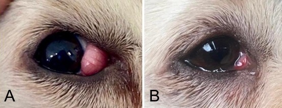
Prolapsed nictitating membrane gland, or "cherry eye," is a common ocular condition in dogs, characterized by protrusion of the third eyelid gland due to weakened connective tissue, often causing inflammation and infection. A 1-year-old, 11 kg male Kintamani dog presented with a persistent reddish mass protruding from the corner of the right eye for three months. Clinical examination, history, and physical findings confirmed nictitating membrane prolapse (cherry eye), with a favorable prognosis. Cherry eye occurs when the gland of the third eyelid prolapses from its normal position, forming a swollen red mass at the medial canthus. Surgical correction was performed using the Morgan's pocket technique. The dog was premedicated with atropine sulfate, and anesthesia was induced using xylazine and ketamine. The prolapsed gland was repositioned between the two incision lines with gentle downward pressure and then sutured using 4-0 PGA (Assucryl®) in a simple continuous pattern. Postoperative care included antibiotic and anti-inflammatory eye drops (Cendo Xitrol®: Neomycin Sulfate, Polymyxin B Sulfate, Dexamethasone) and oral meloxicam for analgesia. By day 13 post-surgery, the eye had fully recovered, with no signs of recurrence or complications.
Dewangan R, Sharda R, Kalim MO, Panchkhande N, Sidar SK, Sahu D. 2018. Chery eye in crossbred dog and its surgical management. International Journal of Science, Environment and Technology. 7(1):288-291.
Deveci MZ, İşler CT, Yurtal Z, Altuğ ME, Kirgiz Ö. 2022. Evaluation of Morgan's pocket technique in the treatment of nictitans gland prolapse in dogs. Turkish Journal of Veterinary and Animal Sciences. 44(3):521-527. https://doi.org/10.3906/vet-2001-54 DOI: https://doi.org/10.3906/vet-2001-54
Gelatt KN, Gelatt JJP. 2011. Surgical procedures for the conjunctiva and the nictitating membrane. In: Gelatt KN, Gelatt JP, eds. Veterinary Ophthalmic Surgery. Pennsylvania: Elsevier-Saunders. https://doi.org/10.1016/B978-0-7020-3429-9.00007-9 DOI: https://doi.org/10.1016/B978-0-7020-3429-9.00007-9
Multari D, Perazzi A, Contiero B, De Mattia G, Iacopetti I. 2016. Pocket technique or pocket technique combined with modified or-bital rim anchorage for the replacement of a prolapsed gland of the third eyelid in dogs: 353 dogs. Veterinary Ophthalmology. 19(3):214-219. https://doi.org/10.1111/vop.12286 | PMid:26096380 DOI: https://doi.org/10.1111/vop.12286
Oguntoye CO, Kodie DO, Oni ZO, Oyetayo NS, Eyarefe OD. 2022. Modified Morgan Pocket Technique for cherry eye repair in 31 dogs. Alexandria Journal of Veterinary Sciences. 72(2):55-63. https://doi.org/10.5455/ajvs.75027 DOI: https://doi.org/10.5455/ajvs.75027
Peruccio, C. 2018. Slatter's Fundamentals of Veterinary Ophthalmology. St. Louis: Elsevier.
Utomo EB, Lesmana MA, Rickyawan N, Airlangga GW, Waspada, SD. 2022. Manajemen bedah prolaps membran niktitan pada anjing beagle di DNA Animal Clinic Bogor. Veterinary Biomedical Clinic Journal. 4(2):75-84. https://doi.org/10.21776/ub.VetBioClinJ.2022.004.02.5 DOI: https://doi.org/10.21776/ub.VetBioClinJ.2022.004.02.5
All articles published in this journal are licensed under a Creative Commons Attribution-ShareAlike 4.0 International License (CC BY-SA 4.0).
This license permits use, distribution, reproduction, and adaptation in any medium, including for commercial purposes, provided the original work is properly cited, a link to the license is given, and any changes made are indicated. Any derivative work must be distributed under the same license or a compatible license.
How to Cite