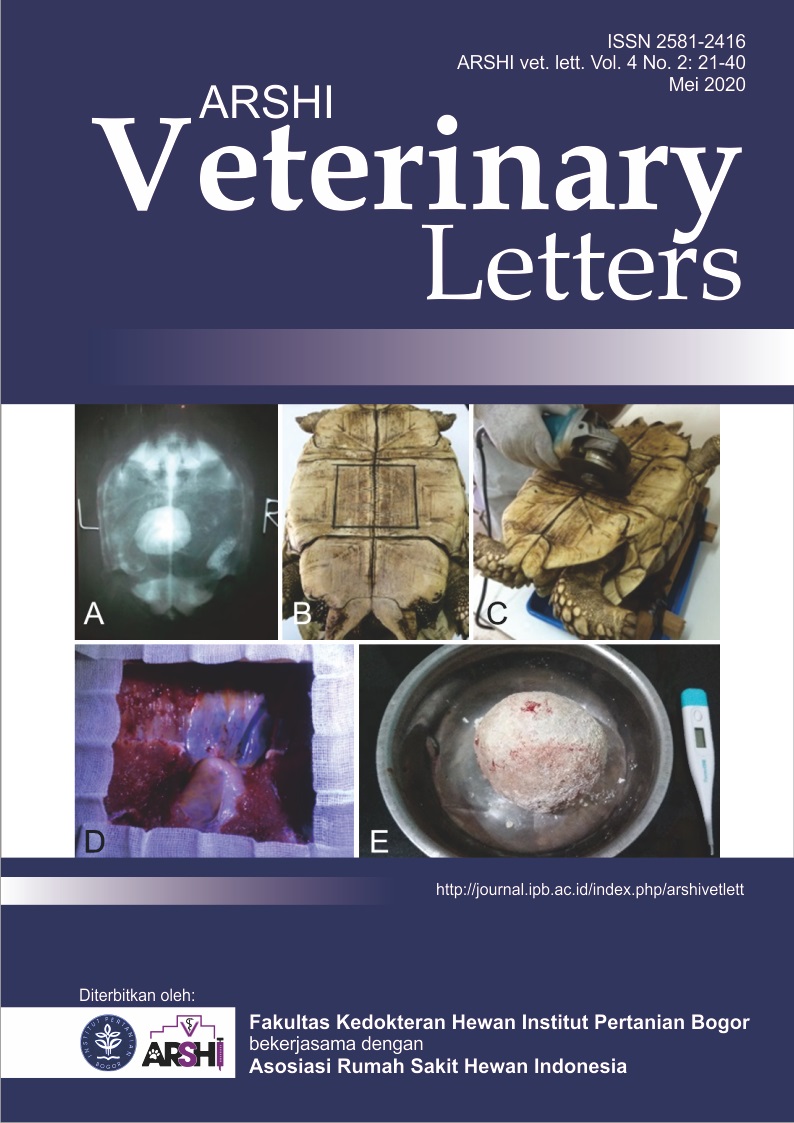
Kuantifikasi opasitas hasil radiografi mesin radiografi analog
Abstract
Citra radiografi suatu objek yang diperoleh dari mesin x-ray analog adalah berupa film yang telah tercetak sebagai suatu gambar dengan gradasi warna hitam, abu dan putih yang disebut sebagai opasitas. Penilaian perubahan opasitas pada film hasil radiografi analog dilakukan dengan bantuan lampu illuminator untuk menampilkan citra dari objek. Identifikasi perubahan opasitas suatu objek dilakukan oleh dokter hewan secara kualitatif dan bersifat sangat subjektif. Tulisan ini menyajikan teknik kuantifikasi sederhana dalam menilai opasitas citra radiografi analog menjadi hasil yang lebih objektif untuk mengurangi subjektifitas. Film radiografi analog yang telah diperoleh selanjutnya diubah menjadi digital secara fotografi menggunakan kamera digital yang ada pada ponsel genggam. Fotografi citra dilakukan di ruangan gelap dengan bantuan lampu illuminator. Foto kemudian dipindahkan ke perangkat komputer untuk diolah dengan perangkat lunak ImageJ. Nilai opasitas suatu area terpilih ditentukan oleh densitas objek berupa gray value pada setiap pixel dalam rentang angka 0-255. Nilai nol untuk citra paling gelap berwarna hitam sebagai radiolucent, sedangkan nilai 255 untuk citra paling terang berwarna putih sebagai radiopaque.
Downloads
References
Geiger M, Blem G, Ludwig A. 2016. Evaluation of imageJ for relative bone density measurement and clinical application. Journal of Oral Health and Craniofacial Science. 1(1):012–021.
Kawai T, Sato K, Yosue T. 2005. Effects of viewing conditions on the detection of contrast details on intraoral radiographs. Oral Radiology. 21(1): 23–29.
Moshfeghi M, Shahbazian M, Sajadi SS, Sajadi S, Ansari H. 2015. Effect of different viewing conditions on radiographic interpretation. Journal of dentistry (Tehran, Iran). 12(11):853-858.
Shi XQ, Sallstrom P, Welander U. 2002. A color coding for radiographic images. Image and Vision Computing. 20(11): 761-767.
Thrall DE, Widner WR. 2013. Chapter I: Radiation Protection and Physics of Diagnostic Radiology, Di dalam Textbook of Veterinary Diagnostic Radiology. 6th ed. Carolina (US): Elsevier Inc.
Copyright (c) 2020 CC-BY-SA

This work is licensed under a Creative Commons Attribution-ShareAlike 4.0 International License.
Authors who publish with this journal agree to the following terms:
1. Authors retain copyright and grant the journal right of first publication with the work simultaneously licensed under a Creative Commons Attribution License that allows others to share the work with an acknowledgement of the work's authorship and initial publication in this journal.
2. Authors are able to enter into separate, additional contractual arrangements for the non-exclusive distribution of the journal's published version of the work (e.g., post it to an institutional repository or publish it in a book), with an acknowledgement of its initial publication in this journal.
3. Authors are permitted and encouraged to post their work online (e.g., in institutional repositories or on their website) prior to and during the submission process, as it can lead to productive exchanges, as well as earlier and greater citation of published work (See The Effect of Open Access).


.jpg)


