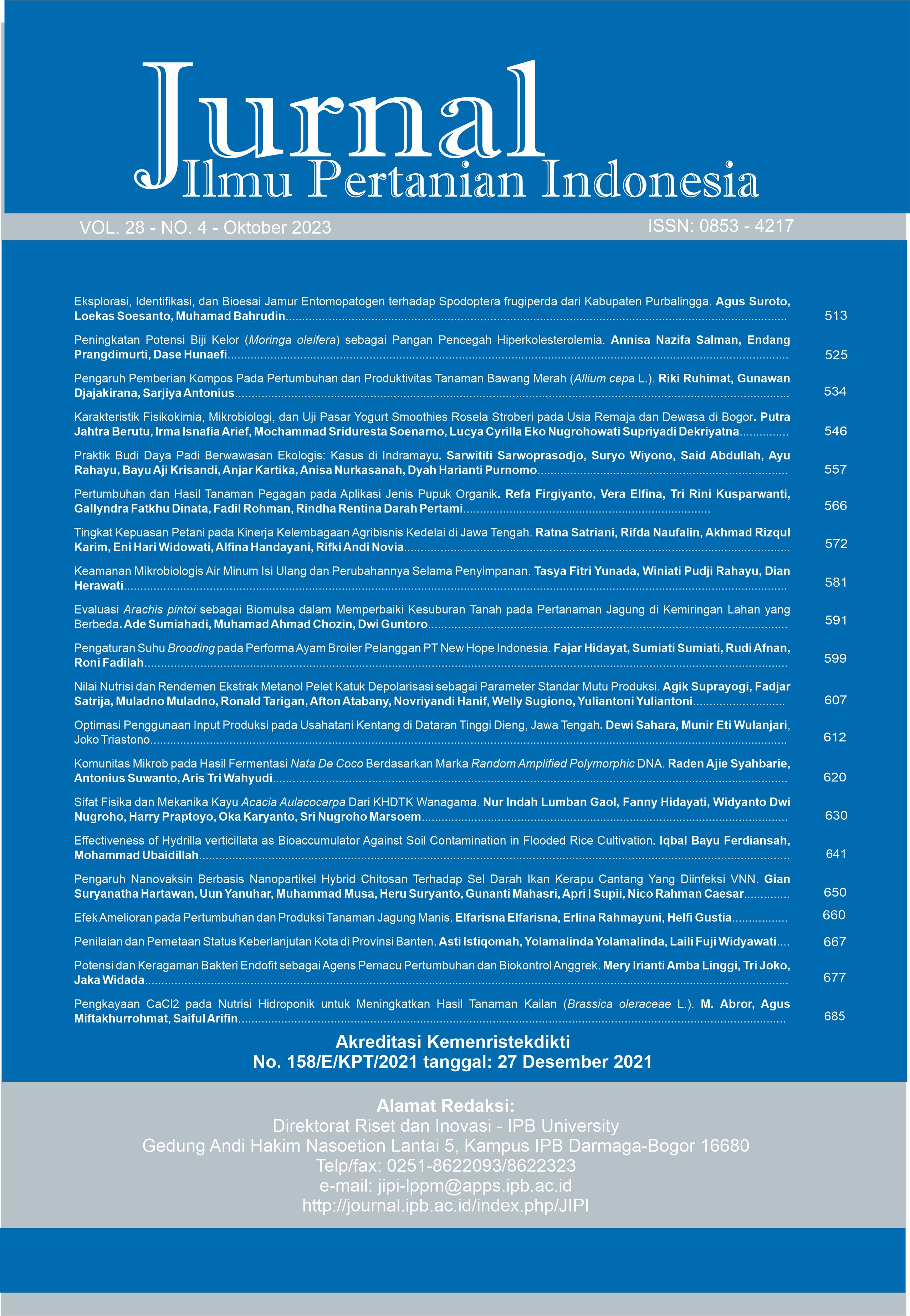
Komunitas Mikrob pada Hasil Fermentasi Nata De Coco Berdasarkan Marka Random Amplified Polymorphic DNA
Abstract
Traditional nata de coco fermentation often results in inconsistent nata thickness. From the producer's perspective, thin nata sheets are detrimental because most fermentation media will be wasted. The main cause of this condition may be that the microbial population in the starter culture is different in each batch. It is necessary to observe the cultured microbial community on various qualities of available thick and thin nata to design a better nata de coco starter culture. This study showed thick nata had more Komagataeibacter intermedius bacteria (pellicle forming) than thin nata. In traditional nata fermentation, K. intermedius always coexists with other microbes from the bacteria and yeast groups. Random Amplified Polymorphic DNA (RAPD) analysis indicated that the genetic diversity of bacteria was higher than that of the yeast group.
Keywords: dendrogram of relationship, fermented food, microbial genetic diversity, nata de coco
Downloads
References
Qureshi AS, Khusk I, Ali CH, Majeed H, Ahmad A. 2017. Production of invertase from Saccharomyces cerevisiae Angel using date syrup as a cost effective carbon source. African Journal of Biotechnology. 16(15): 777–781. https://doi.org/10.5897/AJB2015.15174
Alsulami AMH, Abu-zeid M, Mattar EH, Abo-aba SEM. 2019. Genetic fingerprinting and plasmid content of Acetobacter xylinum strains producing bio-cellulose. World Journal of Medical Sciences. 16(2): 53–58.
Babu KN, Rajesh MK, Samsudeen K, Minoo D, Suraby EJ, Anupama K, Ritto P. 2014. Randomly Amplified Polymorphic DNA (RAPD) and Derived Techniques. Di dalam: Besse P, editor. Molecular Plant Taxonomy. Methods in Molecular Biology (Methods and Protocols). 1115. Totowa (NJ): Humana Press. https://doi.org/10.1007/978-1-62703-767-9_10
Baleiras Couto MM, Eijsma B, Hofstra H, Huis In’T Veld JHJ, Van der Vossen JMBM. 1996. Evaluation of molecular typing techniques to assign genetic diversity among Saccharomyces cerevisiae strains. Applied and Environmental Microbiology. 62(1): 41–46. https://doi.org/10.1128/aem.62.1.41-46.1996
Beneduzi A, Moreira F, Costa PB, Vargas LK, Lisboa BB, Favreto R, Baldani JI, Passaglia LMP. 2013. Diversity and plant growth promoting evaluation abilities of bacteria isolated from sugarcane cultivated in the South of Brazil. Applied Soil Ecology. 63: 94–104. https://doi.org/10.1016/j.apsoil.2012.08.010
Bertsch P, Etter D, Fischer P. 2021. Transient in situ measurement of kombucha biofilm growth and mechanical properties. Food & Function. 12(9): 4015–4020. https://doi.org/10.1039/D1FO00630D
Chávez-Pacheco JL, Martínez-Yee S, Contreras ML, Gómez-Manzo S, Membrillo-Hernández J, Escamilla JE. 2005. Partial bioenergetic characterization of Gluconacetobacter xylinum cells released from cellulose pellicles by a novel methodology. Journal of Applied Microbiology. 99(5):1130–1140. https://doi.org/10.1111/j.1365-2672.2005.02708.x
Fan Y, Huang X, Chen J, Han B. 2020. Formation of a mixed-species biofilm is a survival strategy for unculturable lactic acid bacteria and Saccharomyces cerevisiae in Daqu, a Chinese traditional fermentation starter. Frontiers in Microbiology. 11(February): 1–13. https://doi.org/10.3389/fmicb.2020.00138
Furukawa S. 2015. Studies on formation, control and application of biofilm formed by food related microorganisms. Biosci Biotechnol Biochem. 79(7): 1050–1056. https://doi.org/10.1080/09168451.2015.1018126
Gomes RJ, Borges MF, Rosa MF, Castro-Gómez RJH, Spinosa WA. 2018. Acetic acid bacteria in the food industry: systematics, characteristics and applications. Food Technology & Biotechnology. 56(2): 139–151. https://doi.org/10.17113/ftb.56.02.18.5593
Hammer Ø, Harper DAT, Ryan PD. 2001. PAST : Paleontological Statistics software package for education and data analysis. Palaeontologia Electronica. 4(1): 1–9.
Harrison K, Curtin C. 2021. Microbial composition of SCOBY starter cultures used by commercial kombucha brewers in North America. Microorganisms. 9(5): 1–21. https://doi.org/10.3390/microorganisms9051060
Jagannath A, Manjunatha SS, Ravi N, Raju PS. 2011. The effect of different substrates and processing conditions on the textural characteristics of bacterial cellulose (nata) produced by Acetobacter xylinum. Journal of Food Processing Engineering. 34(3): 593–608. https://doi.org/10.1111/j.1745-4530.2009.00403.x
Kounatidis I, Crotti E, Sapountzis P, Sacchi L, Rizzi A, Chouaia B, Bandi C, Alma A, Daffonchio D, Mavragani-Tsipidou P, et al. 2009. Acetobacter tropicalis is a major symbiont of the olive fruit fly (Bactrocera oleae). Applied and Environmental Microbiology. 75(10): 3281–3288. https://doi.org/10.1128/AEM.02933-08
Lebaron P, Ghiglione J-F, Fajon C, Batailler N, Normand P. 1998. Phenotypic and genetic diversity within a colony morphotype. FEMS Microbiol Letters. 160: 137–143. https://doi.org/10.1111/ j.1574-6968.1998.tb12903.x
Marchesi JR, Sato T, Weightman AJ, Martin TA, Fry JC, Hiom SJ, Wade WG. 1998. Design and evaluation of useful bacterium-specific PCR primers that amplify genes coding for bacterial 16S rRNA. Applied and Environmenta Microbiology. 64(2): 795–799. https://doi.org/10.1128/AEM.64.2.795-799.1998
Moore E, Arnscheidt A, Krüger A, Strömpl C, Mau M. 2004. Simplified protocols for the preparation of genomic DNA from bacterial cultures. Di dalam: Molecular Microbial Ecology Manual. Ed ke-2. Dordrecht (NL): Kluwer Academic Publishers.
Muchtar L, Rachmania Mubarik N, Suwanto A. 2017. Konsistensi produksi nata dalam media fermentasi yang mengandung hidrolisat ubi kayu. Jurnal Teknologi Industri Pertanian. 27(2): 217–227. https://doi.org/10.24961/j.tek.ind.pert.2017.27.2.217
Pavel AB, Vasile CI. 2012. PyElph - a software tool for gel images analysis and phylogenetics. BMC Bioinformatics. 13(9): 1–6. https://doi.org/10.1186/1471-2105-13-9
Sanders ER. 2012. Aseptic laboratory techniques: plating methods. Journal of Visualized Experiments. 63: 1-18. https://doi.org/10.3791/3064-v
Seumahu CA, Suwanto A, Hadisutanto D, Thenawijaya Suhartono M. 2007. The dynamics of bacterial communities during traditional nata de coco fermentation. Microbiology Indonesia. 1(2): 65–68. https://doi.org/10.5454/mi.1.2.4
Smid EJ, Lacroix C. 2013. Microbe–microbe interactions in mixed culture food fermentations. Current Opinion in Biotechnology. 24(2): 148–154. https://doi.org/10.1016/j.copbio.2012.11.007
Steensels J, Gallone B, Voordeckers K, Verstrepen KJ. 2019. Domestication of industrial microbes. Current Biology. 29(10): 381–393. https://doi.org/10.1016/j.cub.2019.04.025
Trček J, Ramuš J, Raspor P. 1997. Phenotypic characterization and RAPD-PCR profiling of Acetobacter sp. isolated from spirit vinegar production. Food Technology & Biotechnology. 35(1): 63–67.
Trček J, Raspor P. 1999. Molecular characterization of acetic acid bacteria isolated from spirit vinegar. Food Technology & Biotechnology. 37(2): 113–116.
Valera MJ, Torija MJ, Mas A, Mateo E. 2015. Acetic acid bacteria from biofilm of strawberry vinegar visualized by microscopy and detected by complementing culture-dependent and culture-independent techniques. Food Microbiology. 46: 452–462. https://doi.org/10.1016/j.fm.2014.09.006
White T, Bruns T, Lee S, Taylor J. 1990. Amplification and direct sequencing of fungal ribosomal RNA genes for phylogenetics. Di dalam: Innis MA, Gelfand DH, Sninsky JJ, White TJ, editor. PCR Protocols: a guide to methods and applications. San Diego (CA): Academic Press, https://doi.org/10.1016/B978-0-12-372180-8.50042-1
Wongjiratthiti A, Yottakot S. 2017. Utilisation of local crops as alternative media for fungal growth. Pertanika Journal of Tropical Agricultural Science. 40(2): 295–304.
Yamada Y, Yukphan P, Vu HTL, Muramatsu Y, Ochaikul D, Tanasupawat S, Nakagawa Y. 2012. Description of Komagataeibacter gen. nov., with proposals of new combinations (Acetobacteraceae). The Journal of General and Applied Microbiol. 58(5): 397–404. https://doi.org/10.2323/jgam.58.397
This journal is published under the terms of the Creative Commons Attribution-NonCommercial 4.0 International License. Authors who publish with this journal agree to the following terms: Authors retain copyright and grant the journal right of first publication with the work simultaneously licensed under a Creative Commons Attribution-NonCommercial 4.0 International License. Attribution — You must give appropriate credit, provide a link to the license, and indicate if changes were made. You may do so in any reasonable manner, but not in any way that suggests the licensor endorses you or your use. NonCommercial — You may not use the material for commercial purposes.


